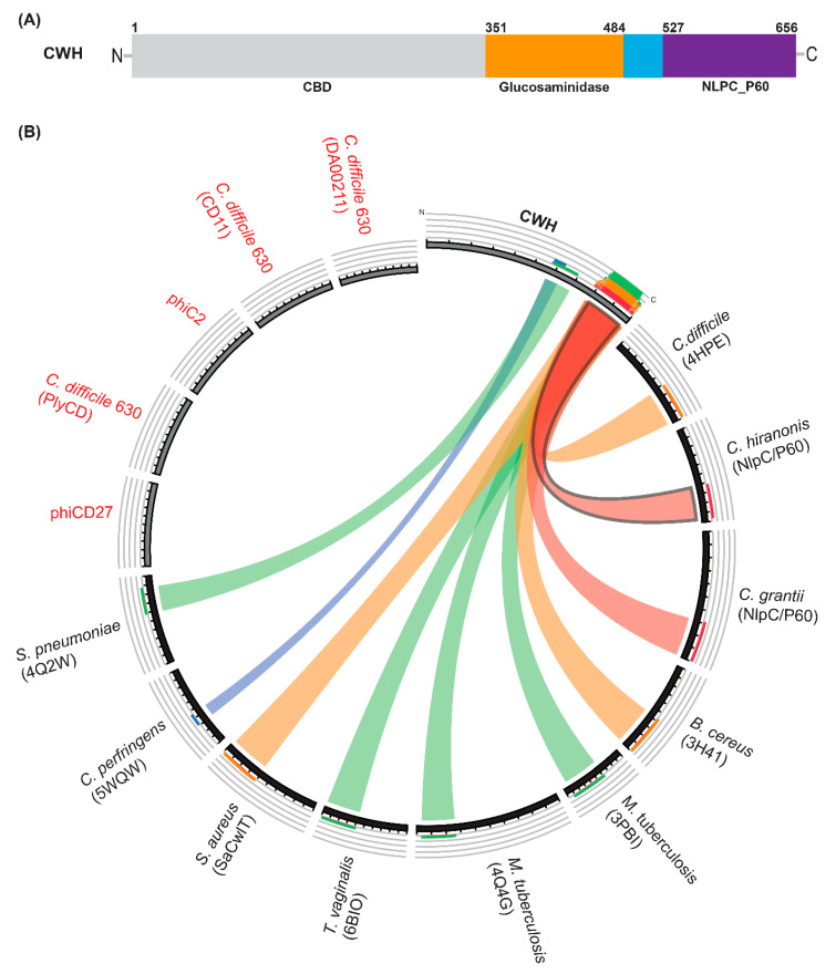
Figure 1.
Modular structure and relationship analysis of cell wall hydrolase (CWH). (A) Schematic presentation of full-length lysin CWH depicting the putative cell wall binding (CBD) domain (light grey), glucosaminidase domain (orange), and Nlp60 (purple). (B) Relationship between CWH and bactericidal enzymes derived from C. difficile phages or prophages and other bacterial species visualized by Circoletto software. The ribbons represent the local alignments produced by BLAST (with an E-value cut off 10−2), and with colours, blue, green, orange, and red representing bit scores of 25%, 50%, 75%, and 100%, respectively. Previously characterised endolysins from C. difficile and phages infecting C. difficile are shown here in red text. Protein names shown in parentheses.

