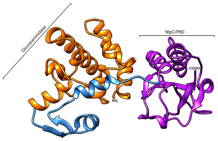
Figure 2.
Homology modelled structure of the CWH351—656. The 3D structure of the protein is represented as a cartoon, with domain and linkers coloured as follows: orange (glucosaminidase), blue (linker), and purple (Nlp60). The model was generated using the Robetta server and visualized using Chimera software.

