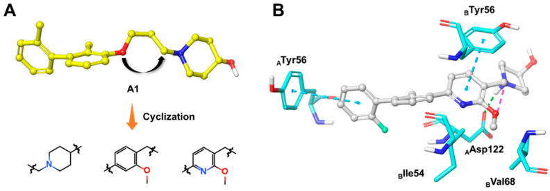
Figure 3.
(A) The optimization strategy of the linker part. (B) Putative binding modes of A4 in the binding pocket of PD-L1 dimer structure (PDB code: 5J89). Dashed lines represent the inter-interaction between A4 and protein, specifically, green ones indicate hydrogen bond, purple ones indicate ionic interaction, and sky-blue ones indicate π–π interaction.

