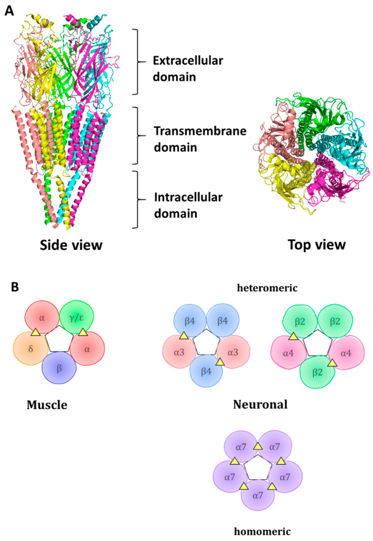
Figure 1.
Representation of the structure of nAChR: (A) Cartoon structure of a muscle-like nAChR with pentameric organization shown through side and top views. Ligand-binding extracellular domain, pore-forming transmembrane domain, and an intracellular domain are shown. PDB accession number is 2BG9. (B) Cartoon representation of assembled muscle and neuronal nAChR subtypes. The pentameric organization of the receptor is shown. The triangles represent the subunit interfaces where endogenous ligands and competitive antagonists bind. In the muscle receptor, a γ-subunit is present in fetal form, while an adult receptor contains the ε- subunit. Two ligand-binding sites include α/δ and α/ε or α/γ interfaces, where the α subunit is considered principal, contributing with loops A, B, and C, whereas δ, ε, and γ are complimentary subunits, contributing with loops D, E, and F to the ligand-binding interface.

