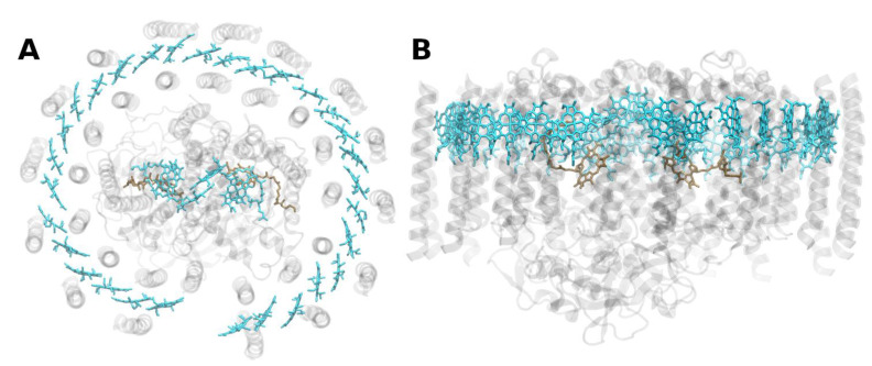
Figure 7.
Structure of the purple-bacterial core-antenna reaction center complex (LH1-RC) from Rhodopseudomonas palustris at 4.8 Å resolution, PDB entry 1PYH [86]. (A) Top view with regard to the bacterial membrane plane, seen from the cytoplasm. (B) Side view with regard to the bacterial membrane plane. The protein backbone is shown in grey. Bacteriochlorophyll a is shown in light cyan, bacteriopheophytin a in brown.

