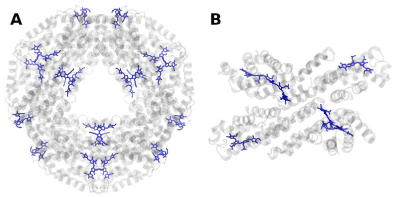
Figure 11.
Structure of phycocyanin from Thermosynechococcus elongatus at 1.35 Å resolution, PDB entry 3L0F [115]. (A) View of the dodecamer along the phycobilisome stacking axis. (B) Side view of a tetrameric unit, perpendicular to the stacking axis. The protein backbone is shown in grey. Phycocyanobilins are shown in blue.

