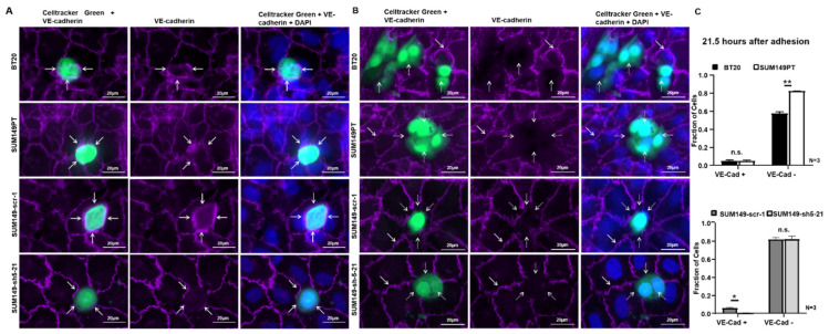
Figure 4.
BT20, SUM149PT, SUM149-scr-1 and SUM149-sh5-21 cells destroy the VE-cadherin-mediated endothelial cell-cell contacts at a late stage of incorporation. The incorporation experiments were stained by immunofluorescence for VE-cadherin (purple) 21.5 h after the start of the experiment. Tumor cells green, 40x magnification and additional + 48% digital zoom. (A) VE-cadherin was present at the contacts between HUVEC and BT20, SUM149PT or SUM149-scr-1 cells, potentially reflecting homophilic interaction between tumor and endothelial cells (white arrows). At the contacts between HUVEC and SUM149-sh5-21 knockdown cells, only endothelial VE-cadherin was visible. Endothelial VE-cadherin between the HUVECs was also present in all images. We interpreted this as an early stage of incorporation; (B) BT20, SUM149PT, SUM149-scr-1 and SUM149-sh5-21 cells disrupted the VE-cadherin mediated HUVEC cell-cell contacts at the site of tumor cell incorporation. BT20, SUM149PT and SUM149-scr-1 cells did not express VE-cadherin on the surface (white dotted arrows); the VE-cadherin staining between adjacent HUVECs was however preserved (white arrows). This might be interpreted as the late stage of the incorporation process; (C) Quantification of incorporation by VE-cadherin immunofluorescence analysis 21.5 h after the start of the experiment. Fraction of tumor cells with endothelial VE-cadherin at junctions to HUVEC (VE-Cad +) and tumor cells that disrupted endothelial VE-cadherin (VE-Cad −). Bars represent standard deviation; n.s. not significant, * p ≤ 0.05; ** p ≤ 0.01; statistical analysis was conducted using two-way ANOVA.

