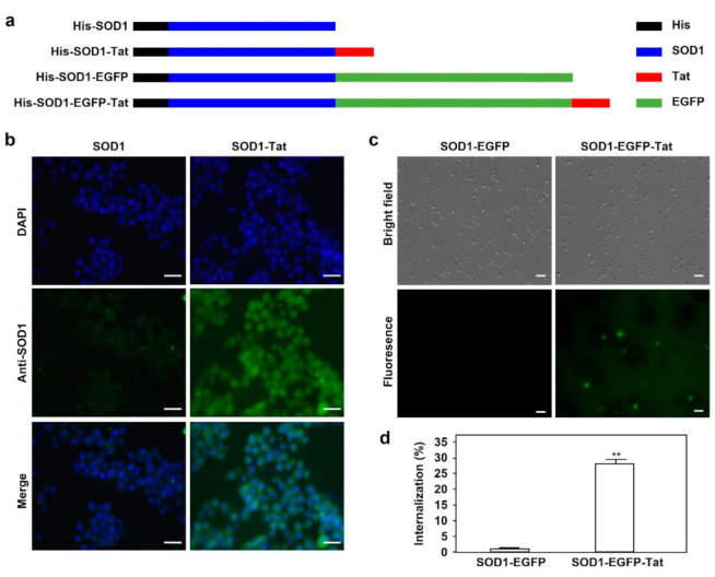
Figure 1.
Tat-mediated cell-penetrating activity of SOD1 fusion proteins. (a). Schematic structures of His-SOD1, His-SOD1-Tat, His-SOD1-EGFP, and His-SOD1-EGFP-Tat fusion proteins expressed in the Rosetta 2(DE3) strain of E. coli. (b). Tat-mediated cell penetration of SOD1 fusion proteins in RAW264.7 cells. RAW264.7 cells were incubated with 5 μΜ SOD1 or SOD1-Tat fusion protein at 37 °C for 1 h. After incubation with a specific anti-SOD1 antibody and the corresponding secondary antibody, the cells were observed for protein internalization by fluorescence microscopy. Bars represent the length of 40 μΜ. (c). Tat-mediated cell penetration of SOD1 fusion proteins in bone marrow cells. Bone marrow cells were isolated from mouse femurs and incubated with 5 μΜ SOD1-EGFP or SOD1-EGFP-Tat fusion protein at 37 °C for 1 h. Tat-mediated protein internalization was examined by fluorescence microscopy. Bars represent the length of 20 μΜ. (d). Quantitative analysis of Tat fusion protein internalization in bone marrow cells. The percentage of EGFP-positive cells in Figure 1c was calculated under a fluorescence microscope. Data are shown as mean ± SD and represent three independent experiments (n = 3). **: p < 0.01, versus SOD1-EGFP group.

