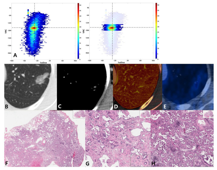
Figure 4.
A 54-year-old woman with lung adenocarcinoma with a 70% acinar and 30% micropapillary pattern. (A) Joint histogram showing a taller than wide shape and compact distribution pattern. (B) Targeted view of the lung window VNC image shows a 24-mm-sized part-solid nodule in the left upper lobe. (C) On conventional enhanced image with mediastinal window, tiny areas of solid portion are seen. (D) On the iodine map, the range of mean iodine value was −51 to 136. (E) PET/CT image shows FDG uptake with SUVmax of 1.6. (F) Photomicrograph (hematoxylin and eosin stain, 10×) shows invasive adenocarcinoma with acinar predominant pattern. High magnification (HE, 100×) shows acinar pattern (G) and micropapillary pattern (H) lung adenocarcinoma.

