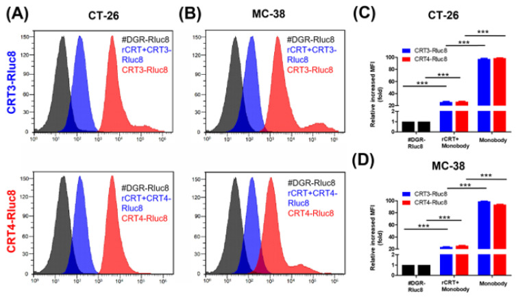
Figure 4.
Monobodies specifically bound to ecto-CRT of cancer cells undergoing ICD. Cancer cells were treated with DOX and recombinant CRT (rCRT), and stained with Rluc8-fused monobodies. After washing, the cells were analyzed for monobody binding by flow cytometry. (A) Flow cytometry of CT-26 cells with exposed ecto-CRT mixed with rCRT. (B) Flow cytometry of MC-38 with exposed ecto-CRT mixed with rCRT. (C,D) Quantitation of (A) and (B). MFI levels of the monobodies bound to ecto-CRT were calculated relative to #DGR-Rluc8. Data are shown as the mean ± standard error (n = 3). *** p < 0.001.

