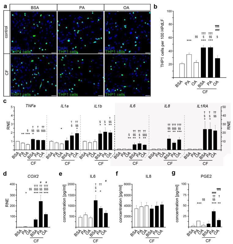
Figure 1.
Palmitic and oleic acid influence the inflammatory response of human periodontal ligament fibroblasts (HPdLF) to compressive force of six hours (CF). (a,b) Analysis of the number of adherent THP1 monocytic cells (green) on HPdLF (blue), stimulated either with palmitic or oleic acid in comparison to BSA control (a). THP1 cells were stained with CellTracker™ and the nuclei of all cells were stained with DAPI. The relative number of THP1 cells is displayed per 100 HPdLF (b). (c,d) Quantitative expression analysis of genes coding for inflammatory markers TNFα, IL1α, IL1β, IL6, IL8, IL1RA (c), and COX2 (d) in fatty acid-cultured HPdLF stimulated with 6 h of compressive force in comparison to BSA controls. Results are normalized to unstimulated BSA controls. (e–g) Analysis of secreted cytokines IL6 (e), IL8 (f), and PGE2 (g) in HPdLF cultures stimulated with palmitic or oleic acid and six hours of compressive force compared to BSA controls. * p < 0.05; ** p < 0.01; *** p < 0.001 in relation to BSA, § p < 0.05; §§ p < 0.01; §§§ p < 0.001 in relation to PA, † p < 0.05; †† p < 0.01; ††† p < 0.001 in relation to OA, # p < 0.05; ### p < 0.001 in relation to BSA+CF, ¶¶¶ p < 0.001 in relation to PA + CF; one-way ANOVA and post hoc test (Tukey). Scale bars: 50 μm in (a). BSA, bovine serum albumin; CF, compressive force; OA, oleic acid; PA, palmitic acid; RNE, relative normalized expression; ~, below detection limit.

