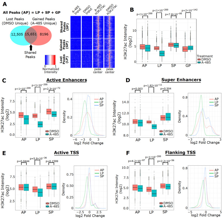
Figure 5.
A-485 preferentially suppresses H3K27ac at enhancers. (A) Venn diagram of the number of H3K27ac peaks detected in four peak types (All Peak (AP), Lost Peak (LP), Gained Peak (GP) and Shared Peak (SP)) in MCF-7 cells treated with A-485 (3 µM) for 24 h (left). Heatmaps depicting normalized H3K27ac peak intensity (at peak center ± 3 kb) in Lost, Gained and Shared peaks are shown (right). (B) Boxplots of H3K27ac peak intensity for DMSO and A-485 in each of the peak types. Boxplots of H3K27ac peak intensity in MCF-7 cells treated with A-485 or DMSO (left) and fold-changes of H3K27ac (right) at active enhancers (C), super enhancers (D), active TSSs, (E) and flanking TSSs, (F) in each of the peak types.

