
Figure 7. Eicosanoids produced during invariant natural killer T (iNKT)–monocyte interactions suppress cord T cells.
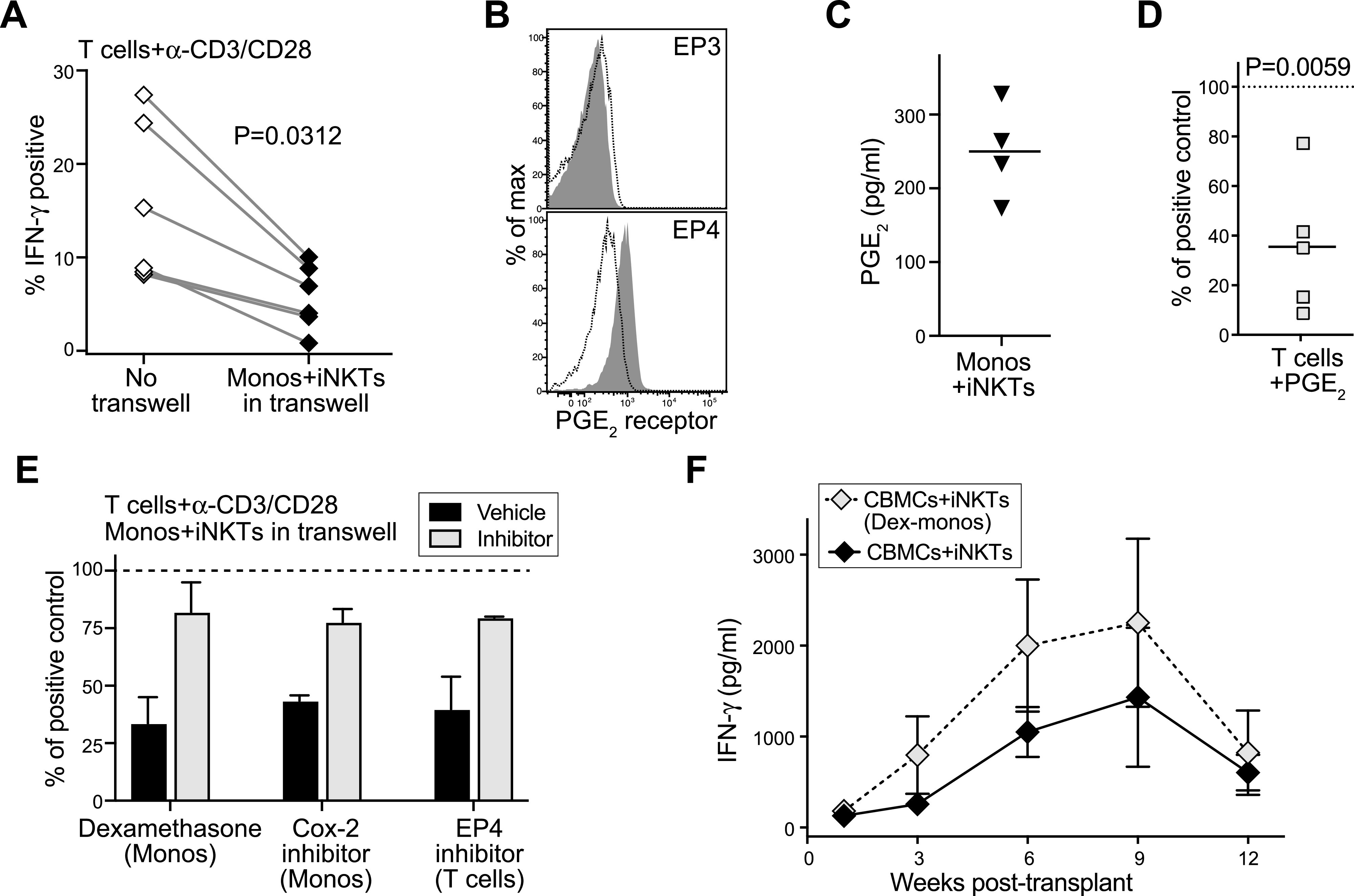
(A) Isolated CD4+ cord blood T cells were cultured for 3–5 d with anti-CD3 and anti-CD28 antibodies in the presence or absence of transwell inserts containing iNKT cells and cord blood monocytes. Plot shows percentage of T cells staining positively for intracellular IFN-γ after PMA/ionomycin stimulation; paired symbols show results from independent experiments. P-value calculated by two-tailed nonparametric paired t test. (B) Flow cytometric staining of cord T cells for PGE2 receptors EP3 and EP4. Filled histograms show staining by specific antibodies; dotted line shows isotype control antibody. (C) iNKT cells and cord blood monocytes were co-cultured for 24 h and PGE2 in the supernatant was quantitated using an enzyme assay. Symbols represent means from independent experiments. (D) Cord blood T cells were cultured for 3–4 d with anti-CD3 and anti-CD28 antibodies in medium containing 500 pg/ml PGE2, then stimulated with PMA/ionomycin. Plot shows the IFN-γ+ T cells as a percent of the response by parallel cultures of cord T cells that were not exposed to PGE2. Symbols show results from independent experiments. P-value calculated by two-tailed one sample t test. (E) Cord blood T cells were cultured for 3–5 d with anti-CD3 and anti-CD28 antibodies in the presence of transwell inserts containing iNKT cells and cord blood monocytes, then stimulated with PMA/ionomycin. Before co-culture, the monocytes were pretreated with dexamethasone (500 ng/ml) or Cox-2 inhibitor (NS-398, 10 μM), or the cord T cells were pretreated with an inhibitor of EP4 (L-161,982, 10 μM). Plot shows IFN-γ production from the transwell co-cultures as a percent of the response by positive control T cells that were cultured with anti-CD3/CD28 alone (dashed line). Bars represent mean ± SEM of two to three independent experiments. (F) NSG mice were transplanted with cord blood mononuclear cells and iNKT cells and blood samples were removed at the indicated times and tested for human IFN-γ by ELISA. Where indicated, the monocytes were isolated and treated with 500 pg/ml dexamethasone, then washed and added back to the cord blood mononuclear cells before injection. Symbols represent mean ± SEM from five mice tested in parallel.