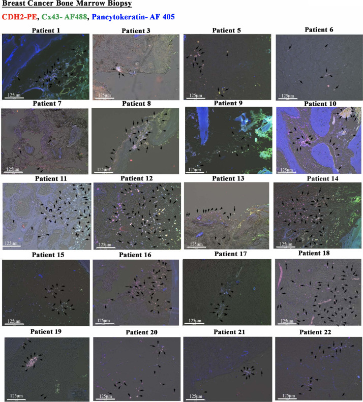
Figure S2. Immunohistochemistry for CDH2 and Cx43 with paraffin sections of BM biopsies from patients with breast cancer.
Tissues were sectioned from paraffin-embedded biopsies of patients diagnosed with breast cancer. The tissues were colabeled with anti–CDH2-PE, anti–Cx43-AF488, and anti–pan-cytokeratin-AF405. The slides were imaged with EVOS FL Auto 2 at 200× magnification. Representative tissues show colocalized Cx43 and CDH2 in pan-cytokeratin+ cells (white spots). Because of the large number of white spots, representative spots are depicted with black arrows.

