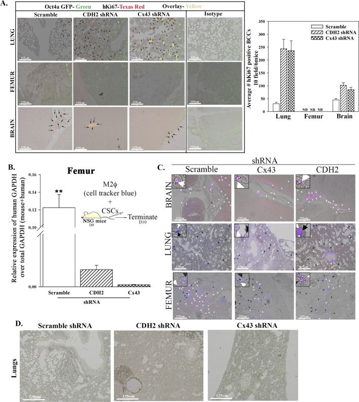
Figure S4. In vivo studies for breast cancer dormancy.
(A) 5 × 10⁵ cancer stem cells (CSCs) from MDA-MB-231-Oct4a-GFP with scramble-RFP, CDH2-shRNA-RFP, or Cx43-shRNA-RFP were injected intravenously into NSG mice as outlined in Fig 4E. At day 10 lung, the femur and brain were harvested. The presence of BC cells in the brain, lungs, and femur was identified with labeling for human Ki67-Texas Red. The green Oct4a-GFP cells were overplayed Ki67-Texas Red–positive cells (yellow cells). Right graph: quantification by ImageJ for Ki67+ BC cells in the femur, lung, and brain, mean of 10 fields/slide for six mice ± SD. (B) 106 CMAC-blue–labeled M2 macrophages were coinjected intravenously with CSCs from MDA-MB-231-Oct4-GFP with scramble-RFP, CDH2-shRNA-RFP, or Cx43-shRNA-RFP. At day 10, the mice were euthanized, and the femur was flushed for RNA isolation. Graph represents the relative expression of human GAPDH over total (human + mouse) GAPDH. (C) The lungs, brain, and femur were imaged for pink cells (blue + RFP). n = 6 mice/group. Tissues were imaged with EVOS FL Auto 2 at magnification of 200×. (D) 5 × 10⁵ CSCs from MDA-MB-231-Oct4-GFP with CDH2-shRNA-RFP, or Cx43-shRNA-RFP were injected intravenously into NSG mice as outlined in Fig 7G. At day 7, the lungs was harvested and imaged for CSCs (yellow cells: GFP+ RFP) using EVOS FL2 at 200× magnification.

