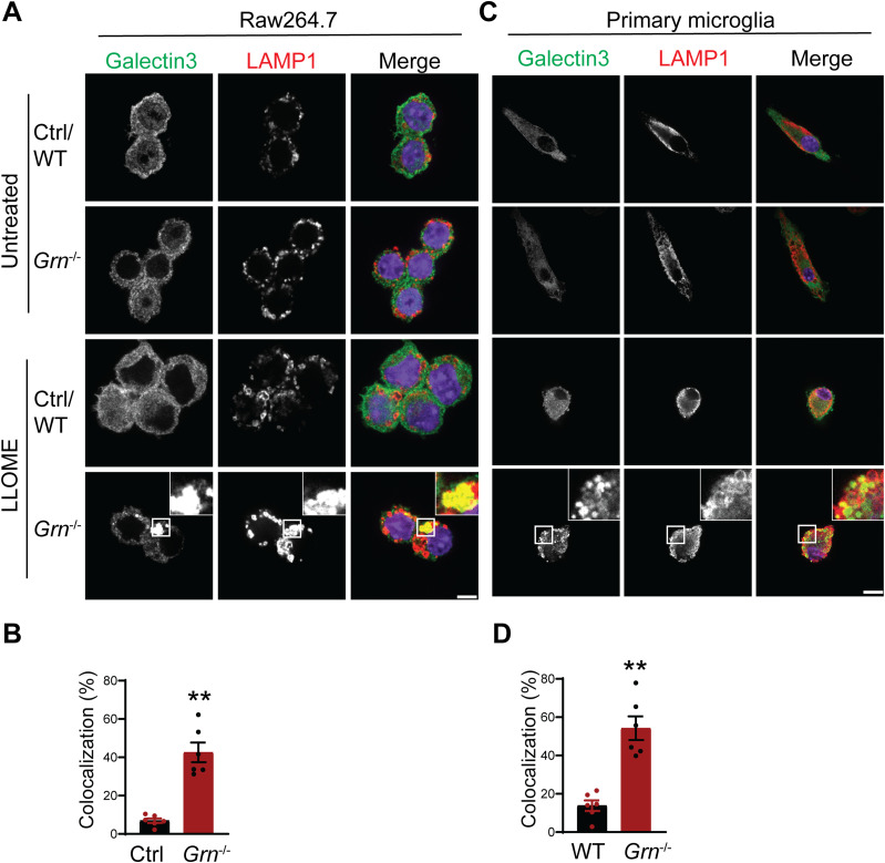
Figure 10. PGRN-deficient macrophage and microglia are more prone to lysosomal membrane leakage.
(A) Representative confocal images of Raw 264.7 cells treated with 500 μM LLOME for 2 h and stained with rat anti-LAMP1 (red) and mouse anti–galectin-3 antibodies (green). Scale bar = 10 µm. (A, B) Manders’ coefficients for colocalization of galectin-3 with LAMP1 for experiment in (A). Mean ± SEM; n = 6, t test, **P < 0.01. (C) Representative confocal images of primary microglia treated with 500 μM LLOME for 2 h and stained with rat anti-LAMP1 (red) and mouse anti–galectin-3 antibodies (green). Scale bar = 10 µm. (C, D) Manders’ coefficients for colocalization of galectin-3 with LAMP1 for experiment in (C). Mean ± SEM; n = 6, t test, **P < 0.01.

