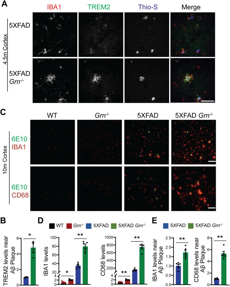
Figure 6. PGRN deficiency leads to increase microglial activation around the Aβ plaques in the 5XFAD mice.
(A) Representative confocal high-resolution images of IBA1, TREM2, and ThioS co-staining in the brain sections from 4.5-mo-old female 5XFAD and 5XFAD Grn−/− mice. Scale bar = 50 µm. (A, B) Quantification of TREM2 signals near Aβ plaques for experiment in (A). Mean ± SEM; n = 3, t test, *P < 0.05. (C) Brain sections from 10-mo-old WT, Grn−/−, 5XFAD and 5XFAD Grn−/− mice were stained with anti-Aβ (6E10) and anti-IBA1 or anti-CD68 antibodies as indicated. Representative images from the cortex region were shown. Scale bar = 100 µm. (C, D) Quantification of IBA1 and CD68 signals in the cortex region for experiment in (C). Mixed female and male mice were used for the study. Mean ± SEM; n = 5, one-way ANOVA, *P < 0.05, **P < 0.01. (C, E) Quantification of IBA1 and CD68 signals near the Aβ plaque in the cortex region for experiment in (C). Mean ± SEM; n = 5, t test, **P < 0.01.

