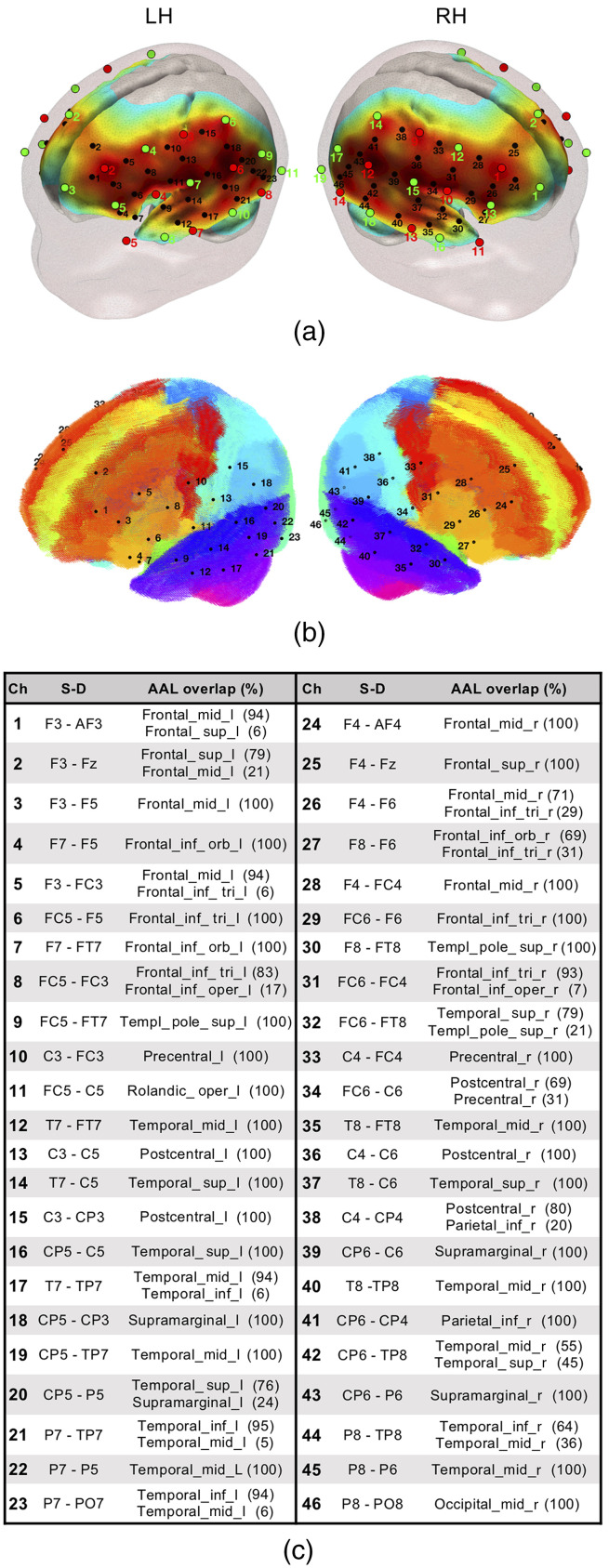
Fig. 1.
(a) fNIRS optode (sources in red and detectors in green) and channel (black) localization in the current experimental setup. The normalized sensitivity profile of this configuration is displayed in a 6-month-old infant head model. (b) Localization of the fNIRS channels in our setup registered to a 6-month-old infant AAL template. (c) Table depicting source–detector distances and the brain labels of our setup based on the on the probabilistic spatial registration of the fNIRS channels to a 6-month-old infant AAL template. Ch, channel and S–D, source–detector pair.

