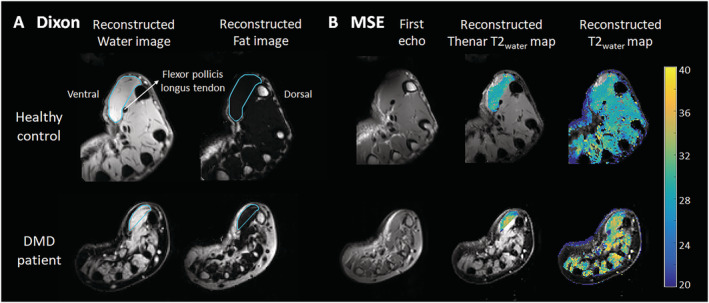
Figure 1.

Dixon and multiecho spin‐echo (MSE) MRI acquisitions and analyses.(A) Example of a Dixon reconstructed water and fat image of a healthy control (HC; 16 years old) and a Duchenne muscular dystrophy (DMD) patient (13 years old). Regions of interest of the thenar muscles were drawn (light blue line) with the dorsal boundary defined as a line on the palmar side of the tendon of the flexor pollicis longus muscle and first metacarpal bone. (B) Example of the first echo from the MSE scan of the same HC and DMD patient and the following reconstructed images: T2water map for voxels within the thenar region of interest and T2water map of all hand muscles. MSE voxels that fitted on the physiological boundaries of the dictionary were excluded.
