
Fig. 4.
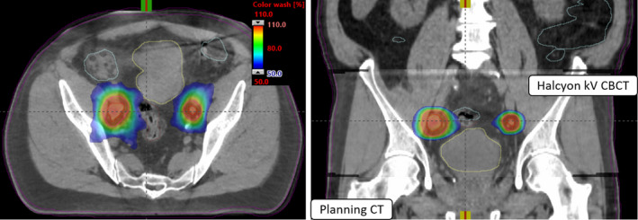
Axial and coronal views of Halcyon kV‐CBCT images (see inset in the coronal view) co‐registered with planning CT images used for image‐guided single‐isocenter (see, cross‐hair) VMAT‐SBRT treatment to both lesions on Halcyon is shown. In addition to anatomical landmarks, the planned dose colorwash (50%–110% isodose cloud) is overlaid for this treatment to demonstrate the delivery accuracy. Halcyon kV‐CBCT images were acquired in the treatment position in free breathing and rigid‐registration was performed via automatic image‐registration mode on Halcyon followed by manually fine‐tuning the registration for better alignment of both nodes via soft‐tissue alignment.