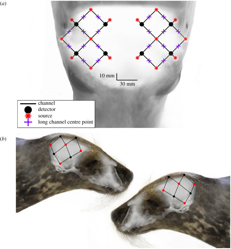
Figure 1.
(a) The optode array configuration overlayed on the head of a seal for anatomical perspective. (b) The anatomically and spatially accurate location of the light sources (red points) and detectors (black points) overlayed on the skull of a juvenile grey seal. Black lines indicate each of twenty 3 cm optode–detector distance channels.

