Figure 2.
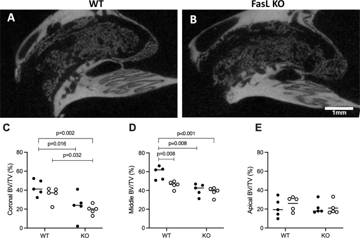
Lack of FasL attenuates regeneration of the extraction socket. Sagittal view of the alveolar socket depicts the WT (A) and FasL KO mice (B). Quantitative analysis of the bone volume per tissue volume (BV/TV) displayed higher amounts of new bone volume in the WT mice in the coronal (C) and middle part (D) of the extraction socket compared to FasL KO mice. The apical region revealed no significant differences in BV/TV between WT and FasL KO mice (E). Statistical analysis was based on Mann‐Whitney U test, P values are given where there was significant differences. The bars show the median and female (black dots) and male mice (white dots) are distributed in the dot plots for the WT and KO group.

