
Figure 6. linc01278 was decreased in proliferative capabilities of vascular smooth muscle cells.
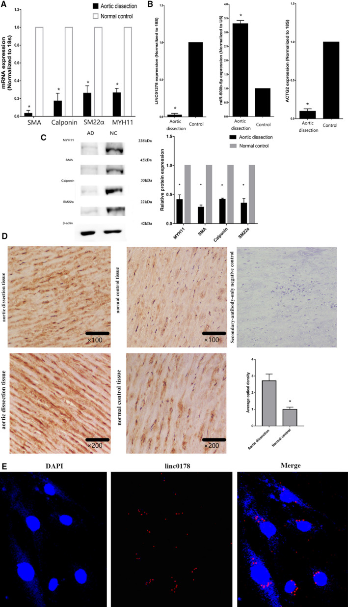
A, Quantitative reverse transcription‐PCR showed the decreased of differentiation marker genes. n=10. *P<0.05. B, Quantitative reverse transcription‐PCR showed the decreased of linc01278 and ACTG2, but miR‐500b‐5p presented with increase. n=10. *P<0.05. C, Decreased differentiation marker genes by Western blot analysis. n=3. *P<0.05. D, Immunostaining showed high expression of Ki‐67 in aortic dissection tissues. n=3. *P<0.05. E, RNA fluorescent in situ hybridization and nucleocytoplasmic separation showed that lnc01278 was mainly expressed in the cytoplasm. AD indicates aortic dissection; DAPI, 4',6‐diamidino‐2‐phenylindole; NC, normal control; and PCR, polymerase chain reaction.