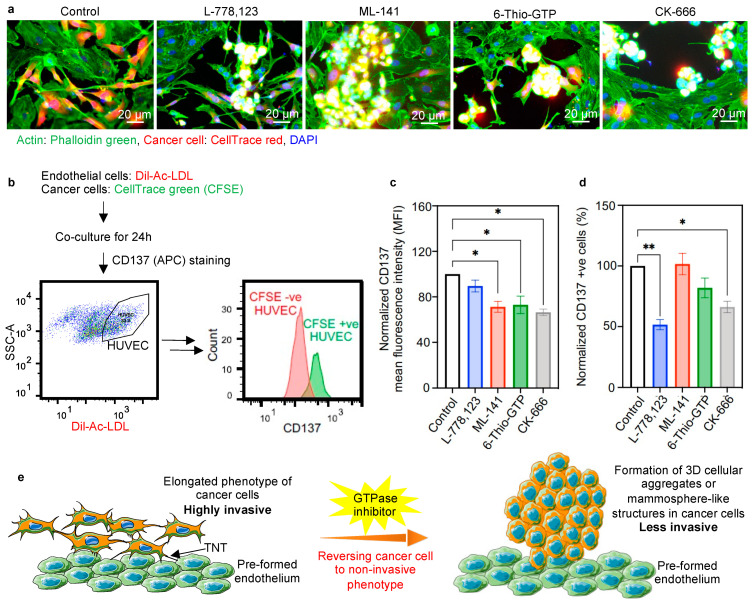
Figure 4.
Reversal of phenotypic transition in the presence of inhibitors. (a) Representative images of co-culture of cancer cells and endothelial cells in the 3D tumor matrix, showing the reversal of the phenotypic transition of cancer cells from a highly invasive elongated phenotype to 3D cellular aggregates or mammosphere-like structures with the effect of inhibitors. Cancer cells (MDA-MB-231) were stained with CellTrace red and co-cultured with unstained endothelial cells. Co-cultures were set up in the absence and presence of inhibitors (10 μM each, 40 μM for CK-666). After 24 h of co-culture, the cells were stained with phalloidin green; nuclei were counterstained with DAPI and imaged under a fluorescence microscope. The mammosphere formation (yellow signal) was observed in the cancer cells in the presence of inhibitors. (b) Experimental design for checking the relative expression of tumor endothelial markers in HUVECs after being involved in nanoscale communication with cancer cells. HUVECs were stained with Dil-Ac-LDL and co-cultured with CFSE stained cancer cells (MDA-MB-231). The histogram showing CFSE +ve HUVECs, i.e., the cells that received cytoplasmic components from cancer cells, exhibited higher CD137 expression than CFSE-ve HUVECs. (c) Representative bar plot showing CD137 mean fluorescence intensity (MFI) of CFSE +ve HUVECs in the presence of GTPase inhibitors. A significant reduction of CD137 MFI was observed that signifies the reduced transfer of information from cancer cells to endothelial cells. (d) Bar plot showing the comparative population of CFSE +ve endothelial cell (Dil-Ac-LDL +ve) with high CD137 expression. A significant reduction in the number of cells expressing high CD137 was observed upon inhibitor treatment. This signifies the reduced transfer of information from cancer cells to endothelial cells. (e) Schematic representation of the effect of inhibitors on the phenotype transition of cancer cells. Metastatic cancer cells adopt an elongated phenotype in the presence of the endothelial layer. The elongated phenotype of cancer cells is able to participate in active communication with endothelial cells, and can easily invade the endothelial layer, resulting in enhanced metastasis. In the presence of GTPase inhibitors, cancer cells return to their original 3D cellular aggregates or mammosphere-like phenotypes, which are less invasive, and which correlate with cancer cells with a reduced chance of invasiveness. * p < 0.05, ** p < 0.01.

