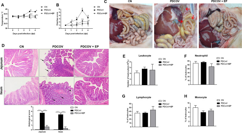
Figure 1.
Effect of EP on PDCoV-associated clinical signs in piglets. Rectal temperature (A) and diarrhea score (B) of piglets that received MEM on days 1–3 (CN), MEM containing a total of 1 × 10⁶ TCID50 of the PDCoV CHN-HN-1601 strain on day 1 and MEM at 8:00 am on days 2–3 (PDCoV), or MEM containing a total of 1 × 10⁶ TCID50 of the PDCoV CHN-HN-1601 strain and EP (2.5 mg/kg body weight) on day 1 and MEM containing EP (2.5 mg/kg body weight) on days 2–3 (PDCoV + EP). Rectal temperature and severity of diarrhea were recorded daily from 0 dpi (the day before PDCoV infection) to 4 dpi. C Macroscopic lesions at 5 dpi. Small intestines of group PDCoV were clearly transparent, thin-walled, and a large amount of yellow fluid accumulated in the intestinal lumen (arrows). D Representative photomicrographs of hematoxylin and eosin-stained jejunal and ileal sections (up) and histological scores of jejunal and ileal tissues (down). Jejunum of group PDCoV showed intestinal villus blunting and rupture, enterocyte attenuation, intestinal epithelial cell shedding (arrows). Ileum of group PDCoV showed mucosal layer edema, atrophy of intestinal glands and basement membrane rupture (arrows). Peripheral blood samples of the indicated groups were collected at 5 dpi. Shown are results for total peripheral blood leukocyte count (E) and the percentage of neutrophils (F), lymphocytes (G) and monocytes (H) of the total number of peripheral blood leukocytes. Data are presented as the mean ± SEM. Mean values at the same time point without a common superscript (a, b, c) differ significantly (P < 0.05). ***P < 0.001. Scale bars, 200 μm.

