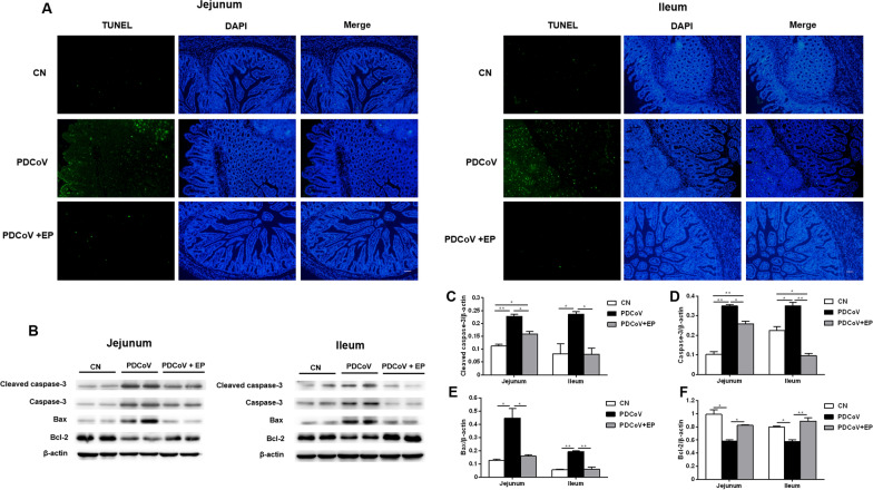
Figure 3.
Effect of EP on PDCoV-induced apoptosis in the small intestine. Intestinal tissue samples of the indicated groups were collected at 5 dpi. A TUNEL assay of paraffin-embedded jejunal and ileal tissues. Green fluorescence represents the TUNEL-positive (apoptotic) cells, and blue fluorescence represents the nuclear distribution. Scale bars, 20 μm. B Western blot analysis of proteins from jejunal and ileal tissues probed with the anti-cleaved caspase-3, anti-caspase-3, anti-Bax and anti-Bcl-2 antibody. C–F. Results are presented as the ratio of target protein band intensity to β-actin band intensity. Data are presented as the mean ± SEM. *P < 0.05; **P < 0.01.

