Figure 8.
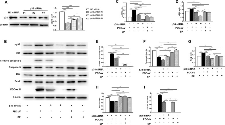
Role of p38 in inhibiting PDCoV replication and alleviating PDCoV-induced apoptosis by EP in LLC-PK1 cells. A The efficiency of p38 siRNA was evaluated by Western blot. LLC-PK1 cells were transfected with the indicated siRNA. At 24 h post-transfection, the expression of p38 was analyzed by Western blot. Results were presented as the ratio of p38 band intensity to β-actin band intensity. B LLC-PK1 cells grown in 6-well plates were transfected with p38 siRNA #1 or NC siRNA for 24 h, and then infected with 2 MOI PDCoV or mock-infected in the absence or presence EP (150 μM). After PDCoV adsorption for 1 h, the cells were further cultured in fresh medium in the absence or presence EP (150 μM). Western blot analysis of proteins from indicated LLC-PK1 cells probed with the anti-p-p38, anti-p38, anti-cleaved caspase-3, anti-caspase-3, anti-Bax, anti-Bcl-2 and anti-PDCoV N antibody. C–I Results are presented as the ratio of target protein band intensity to β-actin band intensity. Data are presented as the mean ± SEM. *P < 0.05; **P < 0.01; ***P < 0.001.

