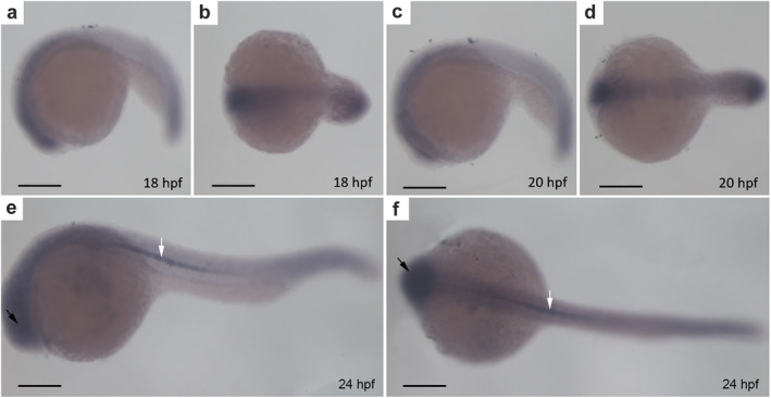
FIGURE 2.

CYLD mainly localizes to the brain and notochord. (a–f) Whole‐mount in situ hybridization of zebrafish embryos. (a–b), (c–d), (e–f) side and top views of zebrafish embryos at 18 hpf, 20 hpf, and 24 hpf, respectively. The black arrows represent the brain of the zebrafish. The white arrows represent the notochord of the zebrafish. Scale bar = 250 μm
