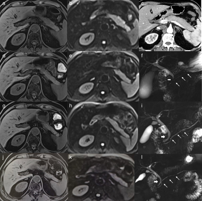
FIGURE 1.

The radiological characteristics and dynamic changes of the patient with severe immune‐related acute pancreatitis (irAP). (a) The T1‐weighted fat‐saturated (T1W FS) image at the onset of severe irAP. (b) The diffusion‐weighted image (DWI) image at the onset of severe irAP. (c) The contrast‐enhanced CT (CECT) image at the onset of severe irAP. (d) The T1w fs image after two weeks of glucocorticoid treatment. (e) The DWI image after two weeks of glucocorticoid treatment. (f) The magnetic resonance cholangiopancreatography (MRCP) showed narrowing of the distal common bile duct (CBD, thick arrow) and main pancreatic duct (MPD, thin arrows), at the onset of severe irAP. (g) The T1W FS image after eight weeks of glucocorticoid treatment. (h) The DWI image after eight weeks glucocorticoid treatment. (i) The MRCP image after eight weeks glucocorticoid treatment. (j) The T1W FS image after 22 weeks of glucocorticoid treatment. (k) The DWI image after 22 weeks of glucocorticoid treatment. (l) The MRCP image after 22 weeks of glucocorticoid treatment
