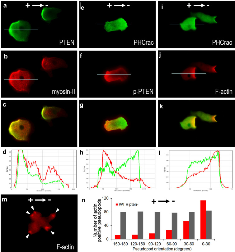
Fig. 7.
Immunocytochemistry staining for PTEN, myosin II, PH-Crac and F-acin in EF. a–d PTEN-GFP and myosin II colocalized at the posterior plasma membrane of the electrotaxing cells. e–h Phospho-PTEN and PHCrac-GFP asymmetrically redistributed to the posterior and anterior membrane of the electrotaxing cells, respectively. i–l PHCrac-GFP and F-actin colocalized at the anterior plasma membrane of the electrotaxing cells. m F-actin leading-edge recruitment was abolished in pten- cells. n Distribution of F-actin positive pseudopods number of EF-treated WT or pten− cells, respectively

