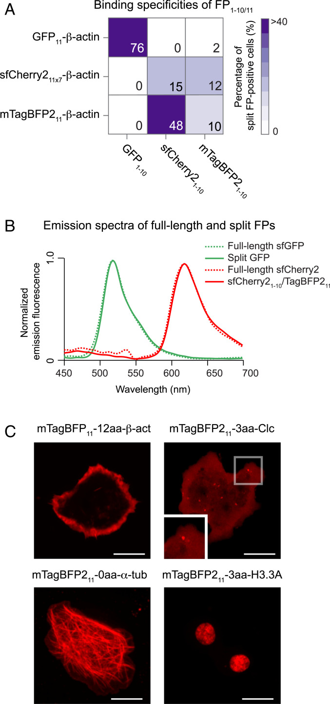
Fig. 3.
Discovery of a bright, red-colored split FP1–10/11 system. (A) Characterizing the binding specificities of three FP1–10/11 pairs (i.e., GFP1–10/11, sfCherry21–10/11 × 7, and mTagBFP21–10/11). We tested each of the FP11 fragments to complement all of the FP1–10 fragments (SI Appendix, Fig. S7). Complementation is indicated as the percentage of fluorescent cells by a color scale and each block’s number. (B) Normalized emission spectra of full-length and split FP variants measured in S2 cells. (C) Fluorescence images of different mTagBFP211 fusions coexpressed with sfCherry21–10 in S2 cells. (Scale bars, 10 μm.)

