Fig. 5.
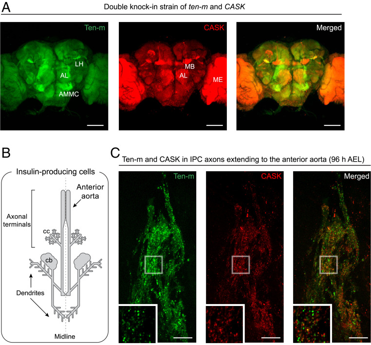
Dual-color endogenous protein tagging in the Drosophila brain. Dual-color fluorescence images of GFP11 × 7-tagged Ten-m and mTagBFP211 × 7-tagged CASK (A) in an adult fly brain, and (C) IPC axonal terminals in a third-instar larval brain. Both GFP1–10 and sfCherry21–10 were (A) panneuronally or (C) IPC-specifically expressed in the double knock-in strain. (B) Schematic drawing of larval IPCs. Their neuronal processes extend to the anterior aorta and the corpora cardiaca (cc) of the ring gland. AL, antennal lobe; AMMC, antenna-mechanosensory and motor center; LH, lateral horn; MB, mushroom body; ME, medulla; cb, cell bodies; and AEL, after egg laying. (Scale bars, 10 μm in C and 100 μm in A.)

