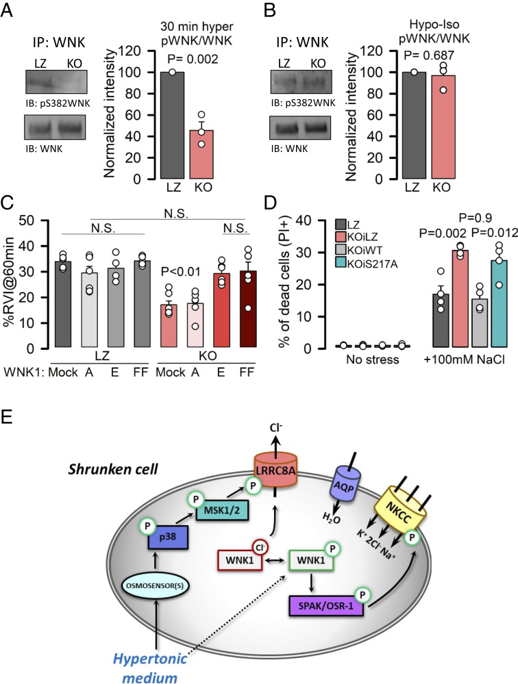
Fig. 4.
LRRC8A triggers WNK activation to promote RVI and cell survival under hypertonicity. (A) Phosphorylation of immunoprecipitated WNK1 obtained from cell lysates of LZ and KO HeLa cells exposed to 30% hypertonic medium. (Left) Western blot of p-S382 and total WNK1; (Right) quantification of phosphorylated WNK1 (normalized to total immunoprecipitated WNK1) after 30-min exposure to hypertonic medium. P values were determined by two-tailed Student’s t test (n = 3). (B) Western blot and quantification of p-S382 WNK1 of immunoprecipitates obtained from cell lysates of LZ or KO HeLa cells (n = 3) in an isotonic medium after exposure to 30% hypotonic medium (as described in Fig. 3G). (C) Mean (±SEM; n = 4 to 7) RVI (%) calculated at 60 min in LZ or KO HeLa cells overexpressing mock, WNK1-S382A (A), WNK1-S382E (E), or WNK1-L369F/L371F (FF). P values were determined by all pairwise one-way ANOVA followed by Holm–Sidak post hoc test. P < 0.01 only when comparing KO mock or KO WNKA with any other condition. (D) PI staining (to monitor cell death) of KO cells expressing WT or S217A mutant LRRC8A under an inducible promoter (DOX). (E) Model of the mechanism that links the p38/MSK1 phosphorylation pathway with the WNK1–NKCC1 axis via LRRC8A to promote RVI and cell survival in response to hypertonic environments.

