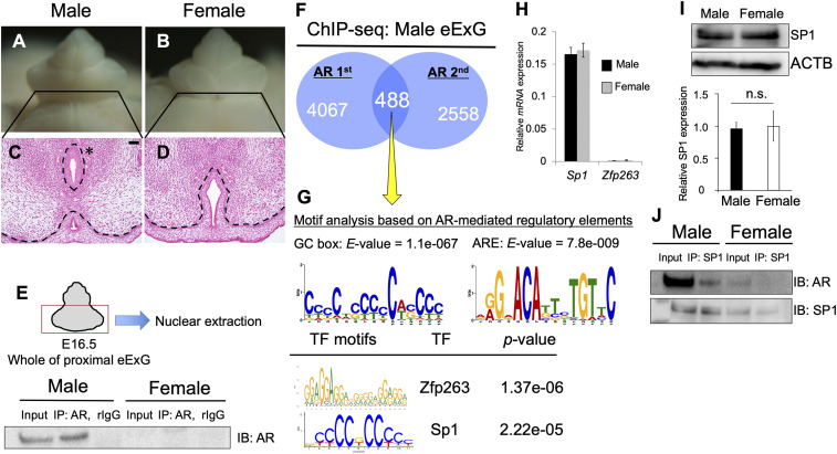
Fig. 1.
Analysis of the regulatory genes for AR transcriptional activity. (A–D) Histological analysis of male and female eExG at E16.5. The asterisk indicates male-type urethra. The urethral plate epithelium remained in female eExG. (Scale bar, 50 μm.) (E) Purified nuclear fraction of whole proximal eExG was utilized for immunoprecipitation with anti-AR or normal rabbit IgG followed by Western blotting with AR antibody. (F) ChIP-seq derived AR cistrome shown as Venn diagram. (G) Motif enrichment analyses from 488 peaks based on AR ChIP-seq (yellow arrow), followed by TOMTOM and MEME analysis. The E-value was utilized to detect significant motif from these analyses. TF, transcription factor. (H) Quantitative real-time PCR from eExG urethral bilateral region mRNA was performed to detect the expression levels of Sp1 and Zfp263 normalized with levels of Gapdh. (I) The levels of SP1 protein normalized by levels of ACTB was detected by Western blotting. Three independent biological replicates were performed. (J) Purified nuclear fraction of whole proximal male and female eExG was utilized for immunoprecipitation with anti-SP1 followed by Western blotting with AR or SP1 antibody. Dashed lines, epithelial–mesenchymal border. n.s. indicates not significant, P > 0.05.

