Fig. 4.
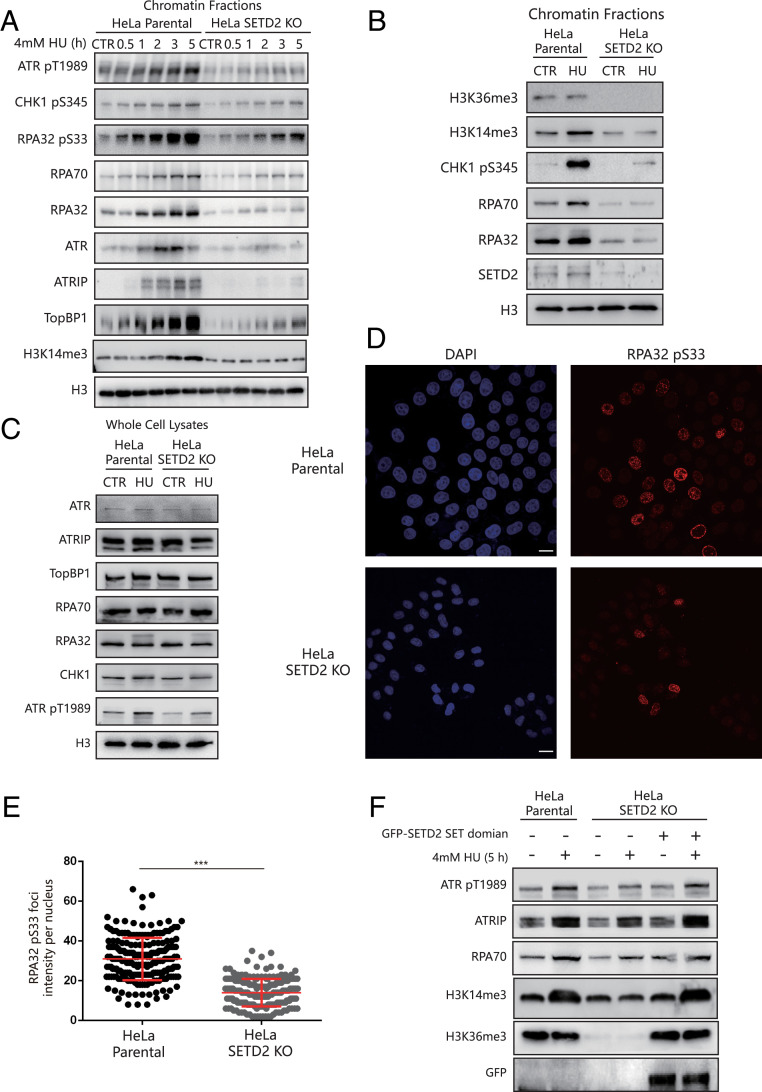
SETD2 depletion impairs ATR activation. (A) HeLa parental cells or SETD2-KO HeLa cells were treated with 4 mM HU for the indicated times, and the chromatin fractions were analyzed by Western blotting. “CTR” indicates the cells without HU treatment. (B) HeLa parental cells or SETD2-KO HeLa cells were treated with 4 mM HU for 5 h, and the chromatin fractions were analyzed by Western blotting. “CTR” indicates the cells without HU treatment. (C) HeLa parental cells or SETD2-KO HeLa cells were treated with 4 mM HU for 5 h, and the whole-cell lysates were analyzed by Western blotting. “CTR” indicates the cells without HU treatment. (D) HeLa parental cells or SETD2-KO HeLa cells were treated with 4 mM HU for 5 h. The cells were then fixed and stained with an anti-RPA32 pS33 antibody (red). (Scale bar, 20 μm.) (E) A statistical analysis of D. The RPA32 pS33 foci per nucleus were counted from a minimum of 200 cells. The data represent the means ± SD, ***P < 0.001 (Student’s t test). (F) SETD2-KO HeLa cells were transfected with or without a vector expressing a GFP-tagged SETD2-SET domain for 60 h. Then, HeLa parental cells or SETD2-KO HeLa cells were treated with or without 4 mM HU for an additional 5 h. The chromatin fractions were extracted for Western blotting.

