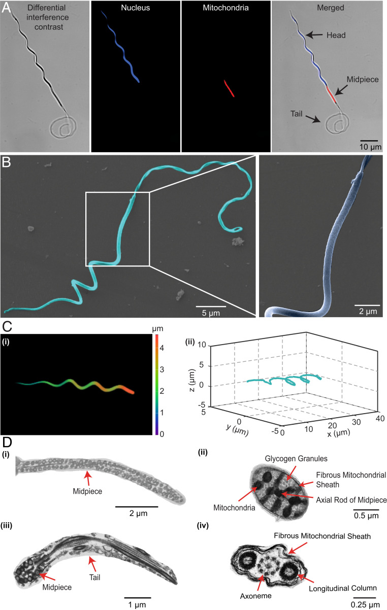
Fig. 1.
The morphology and ultrastructure of Ray sperms. Images of the Ray sperm observed under the fluorescence microscope (A), SEM (B), confocal microscope (C), and TEM (D). The nuclear DNA stained by Hoechst is blue, and the mitochondria stained by MitoTracker are red. Due to the difference in flexibility, the head exhibited helical shapes while the tail curved under microscopes. The lumps on the midpiece represent the mitochondria, which provide energy for the propulsion of sperms. The three-dimensional (3D) helical structure of the head is observed under the confocal microscope (C, i), and the 3D reconstruction of the head is shown in C, ii. The ultrastructure of the sperm is illustrated in D: longitudinal sections of the midpiece (D, i) and tail (D, iii), cross sections of the midpiece (D, ii), and tail (D, iv).

