
FIGURE 3.
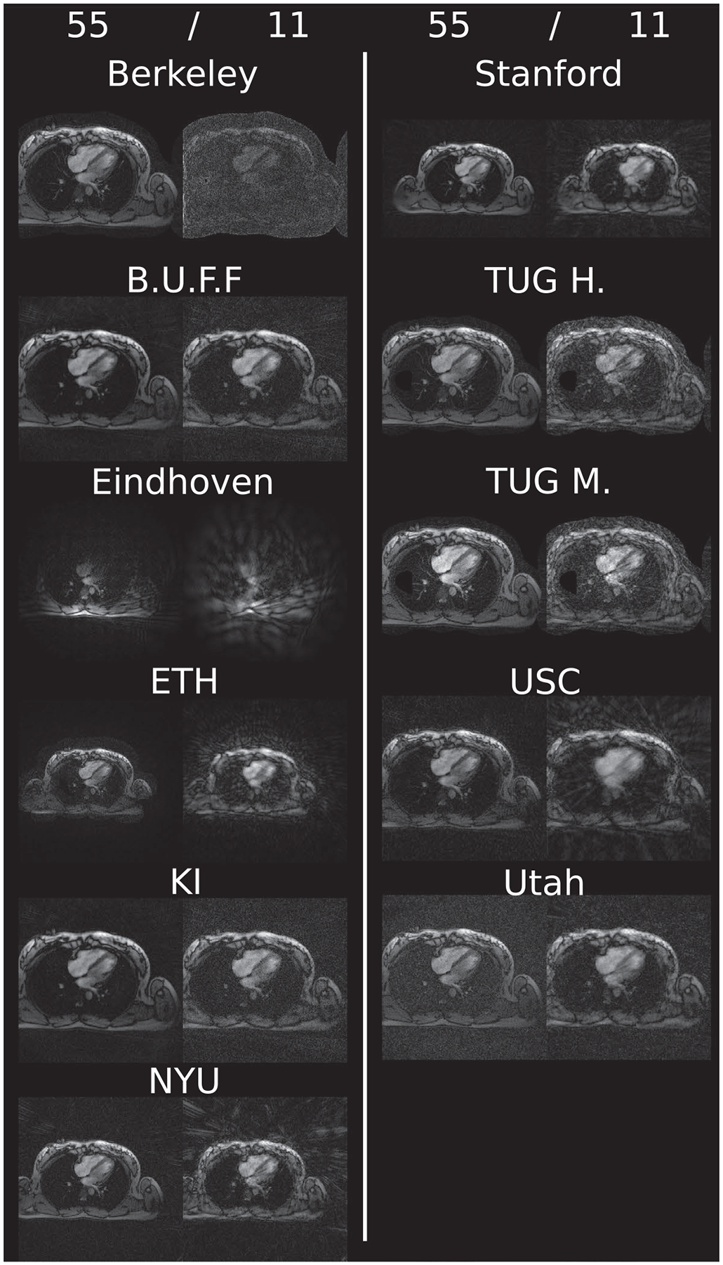
Example reconstruction results for each challenge submission. Shown are results using 55 and 11 spokes of the supplied challenge heart data. Visually observable differences amount to FOV changes as well as image center changes. Intensity variations are not as severe as in the case of brain data. Again, some reconstructions made use of a mask to suppress background signal