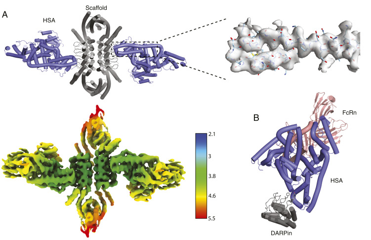
Fig. 5.
Characterization of anti-HSA DARPin assembly in complex with HSA. (A) A cartoon model of scaffold D2-21.8.HSA-C9.v2 and HSA cocomplex docked into a locally refined cryo-EM map obtained from the map below, which is colored by resolution (Å). The density map surrounding the indicated two helices is shown as a transparent gray surface with the model docked. (B) A model of the relative HSA-binding positions of the DARPin and FcRn is built by superposition of this cryo-EM structure (DARPin scaffold + HSA) and an existing crystal structure of HSA with FcRn (Protein Data Bank 4K71). Several hydrophobic DARPin residues involved in binding are shown as sticks.

