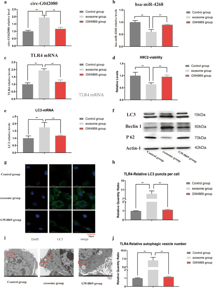
Fig. 6.
Effects of exosomes from MM patients on H9C2 cells. A The level of circ-G042080 was significantly increased in the exosome group (P < 0.01). After blocking the exosomes with GW4869, the level of circ-G042080 decreased. B The level of hsa-miR-4268 was significantly increased in the exosome group (P < 0.01) and then decreased after blocking exosomes with GW4869. C, A The level of TLR4 was significantly increased in the exosome group (P < 0.01) and then decreased after blocking exosomes with GW4869. D CCK-8 assay results showed that the proliferation rate of H9C2 cells in the exosome group was significantly reduced (P < 0.01) and that the cell proliferation rate increased after the exosomes were blocked with GW4869. E The level of LC3 was significantly increased in the exosome group (P < 0.01) and then decreased after blocking exosomes with GW4869. F WB results showed increased LC3-II/I and Beclin1 levels and a decreased P62 level in the exosome group compared with the control group. G Immunofluorescence results showing the LC3 expression level in H9C2 cells. H Compared with that in the control group, the LC3 expression level in the exosome group was significantly increased (P < 0.01). I Observation of the number of autophagic vesicles in H9C2 cells via electron microscopy. The red marks from left to right indicate mitochondria, autophagic vesicles, autophagic lysosomes, mitochondria, and mitochondria. J The results showed easily visible autophagic vacuoles in the exosome group, and the number was obviously greater than that in the control group

