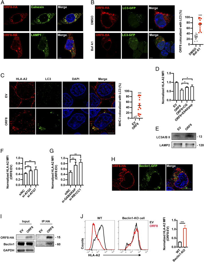
Fig. 5.
ORF8 mediates MHC-Ι degradation through autophagy pathway. (A) Localization of SARS-CoV-2 ORF8-HA (red) relative to CALNEXIN (green, Top) or LAMP1 (green, Bottom). ORF8-HA–expressing plasmid was transfected into HEK293T cells. At 24 h after transfection, colocalization was visualized by confocal microscopy (Scale bars, 5 μm). (B) Localization of SARS-CoV-2 ORF8 (red) relative to LC3-GFP (green). ORF8-HA– and LC3-GFP–expressing plasmids were cotransfected into HEK293T cells. At 24 h after transfection, colocalization was visualized by confocal microscopy (Scale bars, 5 μm) (n = 14 to 20 fields). (C) Localization of HLA-A2 (red) relative to LC3 (green). ORF8-HA–expressing plasmids were transfected into HEK293T cells. At 24 h after transfection, colocalization was visualized by confocal microscopy (Scale bars, 5 μm) (n = 14 to 20 fields). (D) GFP- (empty vector, EV) or ORF8-GFP–expressing plasmid was transfected into HEK293T cells. Before harvest, cells were then treated with chloroquine (CQ) (50 μM) and E64d (10 μg/mL) and pepstatin A (pep) (10 μg/mL) for 6 h. The HLA-A2 mean fluorescence intensity (MFI) (gated on GFP+ cells) was normalized to GFP group (n = 5). (E) EV- or ORF8-HA–expressing plasmid was transfected into HEK293T cells. Cells were treated with Baf A1 (100 nM) for 16 h before harvest for crude lysosomal fraction. Accumulation of LC3B in lysosomes was analyzed by Western blotting. (F and G) GFP (EV)- or ORF8-GFP–expressing plasmids and the indicated siRNAs were transfected into HEK293T cells. MFI of HLA-A2 (gated on GFP+ cells) was normalized to GFP group (n = 5). (H) Localization of SARS-CoV-2 ORF8-HA (red) relative to Beclin 1-GFP (green) (Scale bars, 5 μm). ORF8-HA– and Beclin 1-GFP–expressing plasmids were cotransfected into HEK293T cells. At 16 h after transfection, colocalization was visualized by confocal microscopy (n = 14 to 20 fields). (I) ORF8 was co-IP with Beclin 1. Empty vector (EV)-, or ORF8-HA–expressing plasmid was transfected into HEK293T cells, respectively. Cells were treated with Baf A1 (100 nM) for 16 h before collected. The cells were collected at 48 h after transfection and treated with cross-linker DSP and co-IP with the anti–HA-tag beads (n = 5). (J) GFP (EV)- or ORF8-GFP–expressing plasmids were transfected into HEK293T cells (WT), or Beclin 1 knockout HEK293T cells. Cells were collected at 48 h after transfection, and HLA-A2 MFI was analyzed by flow cytometry (gated on GFP+ cells) and normalized to GFP (EV) group (n = 5). The data were shown as mean ± SD (error bars). Student’s t test and one-way ANOVA was used. P < 0.05 indicates statistically significant difference; *P < 0.05; **P < 0.01; ***P < 0.001.

