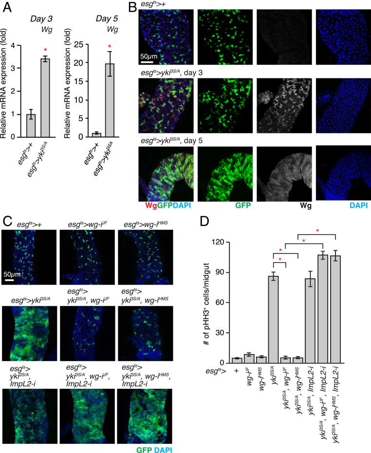
Fig. 1.
Wg is indispensable for the growth of the yki3S/A tumor in the presence of ImpL2. (A) Expression of wg mRNA in midguts. The relative abundance of the wg transcript in esgts>+ or esgts > yki3S/A midguts was determined by qRT-PCR after 3 and 5 d of transgene expression. (B) Immunostaining of Wg in posterior midguts. Transgenes were induced for 3 and 5 d. The cells manipulated by esgts are marked by GFP (green), Wg staining is shown in red, and nuclei are stained with DAPI (blue) in merged images. (Scale bar, 50 µm.) (C) Representative images of posterior midguts. Transgenes were expressed for 5 d. (D) Quantification of pHH3+ cells per midgut. RNAi lines: wg-iJF, JF01257; wg-iHMS, HMS00794; ImpL2-i, 15009R-3. n = 20 (esgts>+), n = 11 (esgts > wg-iJF), n = 11 (esgts > wg-iHMS), n = 22 (esgts > yki3S/A), n = 12 (esgts > yki3S/A, wg-iJF), n = 10 (esgts > yki3S/A, wg-iHMS), n = 12 (esgts > yki3S/A, wg-iJF, ImpL2-i), n = 12 (esgts > yki3S/A, wg-iHMS, ImpL2-i), n = 22 (esgts > yki3S/A, ImpL2-i) biological replicates. Mean ± SEMs are shown. *P < 0.01, two-tailed unpaired Student’s t test compared with control (esgts>+) unless indicated by bracket. See also SI Appendix, Figs. S1–S3.

