Fig. 1.
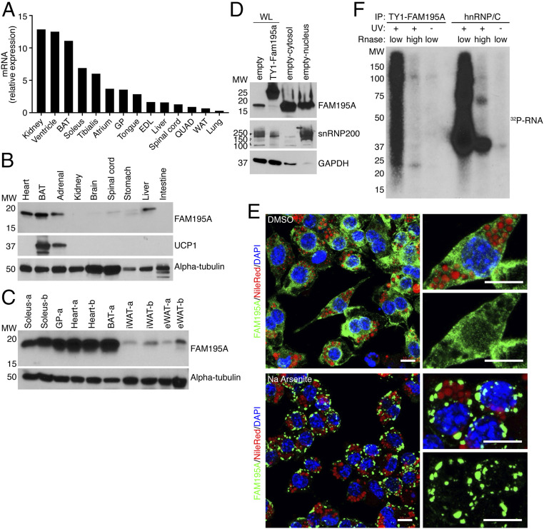
FAM195A is enriched in muscle and BAT. (A) Detection of Fam195a mRNA by qRT-PCR in an adult mouse tissue library; 18S ribosomal RNA was used for normalization. Expression is shown relative to liver. (B) Detection of FAM195A and UCP1 proteins in adult mouse tissue homogenates by western blot analysis. The antibody against alpha-tubulin is used as the loading control. (C) Detection of FAM195A protein levels in different adipose tissue types and different muscle groups; alpha-tubulin protein was used as a loading control. The letters -a and -b represent lysates from two different animals. (D) Western blot analysis showing FAM195A protein in nuclear and cytosolic fractions obtained from differentiated adipocytes. snRNP200 and GAPDH protein are used as controls for cell fractionation efficiency. SVF cell lysate expressing TY1-FAM195A is the control for antibody specificity. (E) Immunofluorescence of FAM195A (green), DAPI for nuclei (blue), and Nile Red for lipid droplets (red). Staining was performed after 30’ incubation with sodium arsenite (500 µM) to induce stress granule formation or dimethyl sulfoxide (DMSO) as control. (Scale bars: 10 µm.) (F) Autoradiograph resolving the FAM195A–RNA complexes after immunoprecipitation (IP) of TY1-epitope–tagged FAM195A in UV cross-linked and uncross-linked adipocytes. As a control, UV cross-linked lysates were treated with high ribonuclease (RNase) concentration. hnRNP/C–RNA complexes were obtained as a control for RNA labeling and UV cross-linking. WL, whole cell lysate; MW, molecular weight; BAT, interscapular brown adipose tissue; EDL, extensor digitorum longus; GP, gastrocnemius plantaris; QUAD, quadricep; WAT, white adipose tissue.

