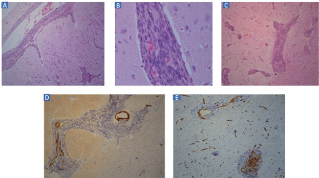
Figure 3.

(A) Perivascular sleeves of spindled to plump cells extending from leptomeningeal vessels into Virchow Robin spaces in cortex (H&E stain, original magnification ×100). (B) High power view of sleeves of spindled to plump cells distending perivascular spaces in deep cortex (H&E stain, original magnification ×400). (C) Perivascular spaces in deep cortex and superficial subcortical white matter expanded by sleeves of spindled to plump cells (H&E stain, original magnification ×100). (D) Focal, weak smooth muscleactin immunolabelling of perivascular cells. Dense staining restricted to vascular media (smooth muscle actin immunostaining, ×100 original magnification). (E) Focal, inconsistent CD34 immunolabelling of perivascular cells (CD34 immunostaining, ×100 original magnification).
