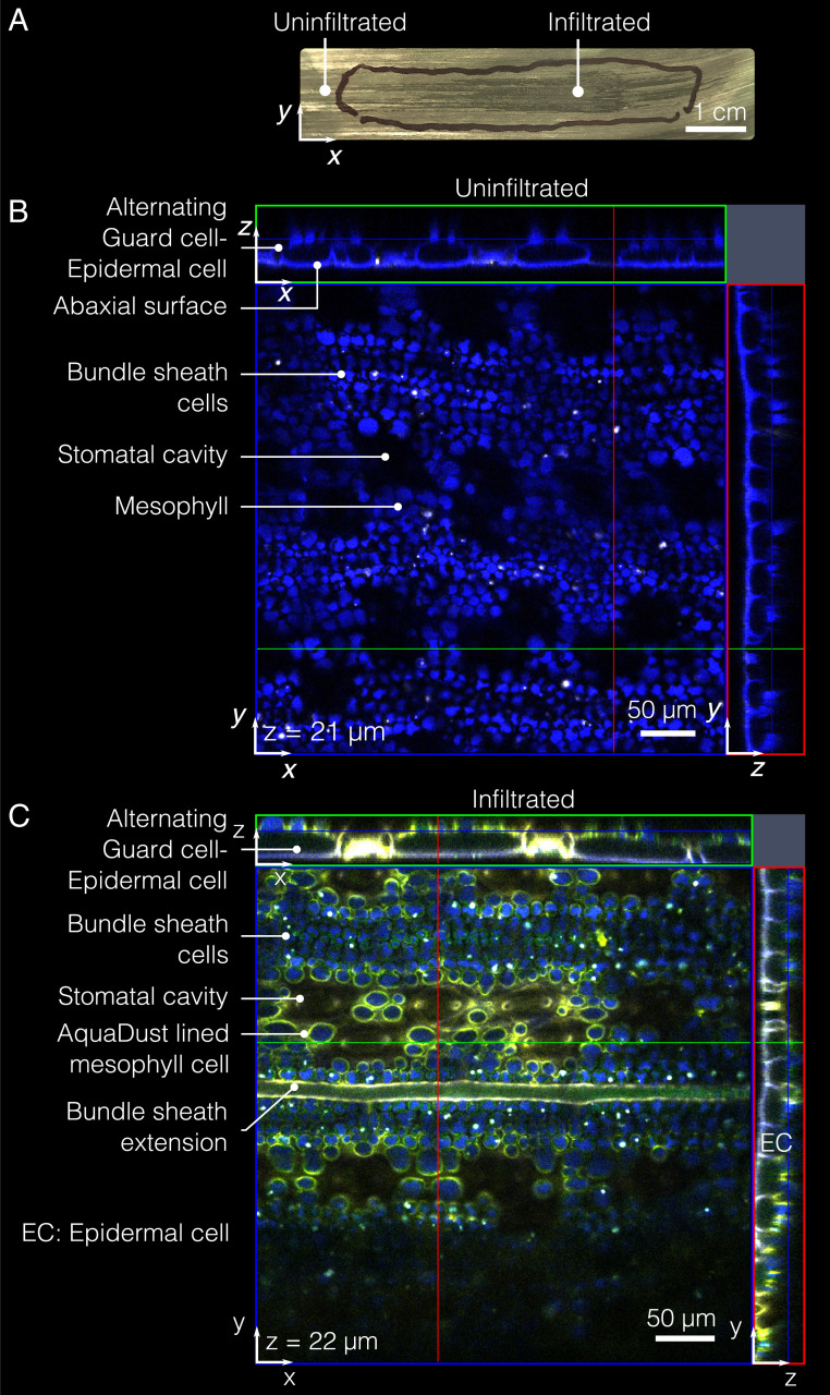
Fig. 2.
AquaDust distribution within mesophyll. (A) Typical infiltration of AquaDust suspension in maize leaf is evident with darkening of infiltrated zone immediately after infiltration; the discoloration dissipates within h as the injected zone re-equilibrates with the surrounding tissue. (Scale bar: 1 cm.) (B) Cytosol and cuticle autofluorescence (blue) from an uninfiltrated maize leaf imaged from the abaxial side using confocal microscope with xz- and yz-planes at locations denoted by green and red lines. (C) Cytosol and cuticle autofluorescence (blue) and AquaDust fluorescence (yellow) as seen from the abaxial side of maize leaf under confocal microscope infiltrated with AquaDust suspension. (See SI Appendix, section S4D for details of preparation and imaging.)

