Fig. 7.
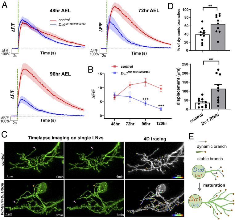
Dα1 mediates neurotransmission at mature LNv synapses. (A and B) Dα1 deficiency leads to a progressive decline in the light-evoked calcium responses during development. Calcium transients were recorded in LNv axonal terminals at different developmental stages. The average trace of calcium transients (A) and the trend line of average peak amplitude (ΔF/F) (B) are shown. The peak amplitude data for 120 h AEL are from Fig. 3C. Significant differences between Dα1 mutants and wild-type controls are found at 96 and 120 h AEL. The shaded area over the average trace represents SEM. The dashed green line indicates the 100-ms light pulse. Sample size n represents the number of larvae tested for each genotype at each time point, n = 9 to 21. Statistical significance is assessed by one-way ANOVA followed by Tukey’s post hoc test. ***P < 0.001. Error bars represent mean ± SEM. (C) Knocking down Dα1 in LNvs leads to elevated dendrite dynamics. Time-lapse imaging of LNv dendritic arbors at the third instar stage reveals the dynamic behavior of individual branches in control (Top) and Dα1 knockdown (Bottom) groups. Representative maximum projected images of single-labeled LNvs are shown. (Left) Two frames, collected at 0 and 4 min in the imaging series, demonstrating representative extension (yellow arrowhead) and retraction (pink arrowhead) events. (Right) 4D tracings of branch terminals depicting the path of dynamic filopodia over the 10-min recording session. Scale bars are as indicated. (D) Quantification of dendrite dynamics. LNv-specific knockdown of Dα1 results in an increased percentage of dynamic branches (Left) as well as increased total displacement of branches (Right). Sample size n represents the number of larvae. n = 10 in both groups. Statistical significance is assessed by Student’s t test. **P < 0.01. Error bars represent mean ± SEM. (E) A schematic diagram illustrating the contributions of Dα1 and Dα6 in LNv dendritic development. Immature LNvs are characterized by a high expression of Dα6, a low expression of Dα1, and highly motile dendrite branches. During larval development, Dα6 and Dα1 is either down- or up-regulated, respectively, accompanying synapse maturation as well as dendrite stabilization and expansion.

