
Fig 1. Evaluation of [18F]florbetapir lung uptake and CT lung density values.
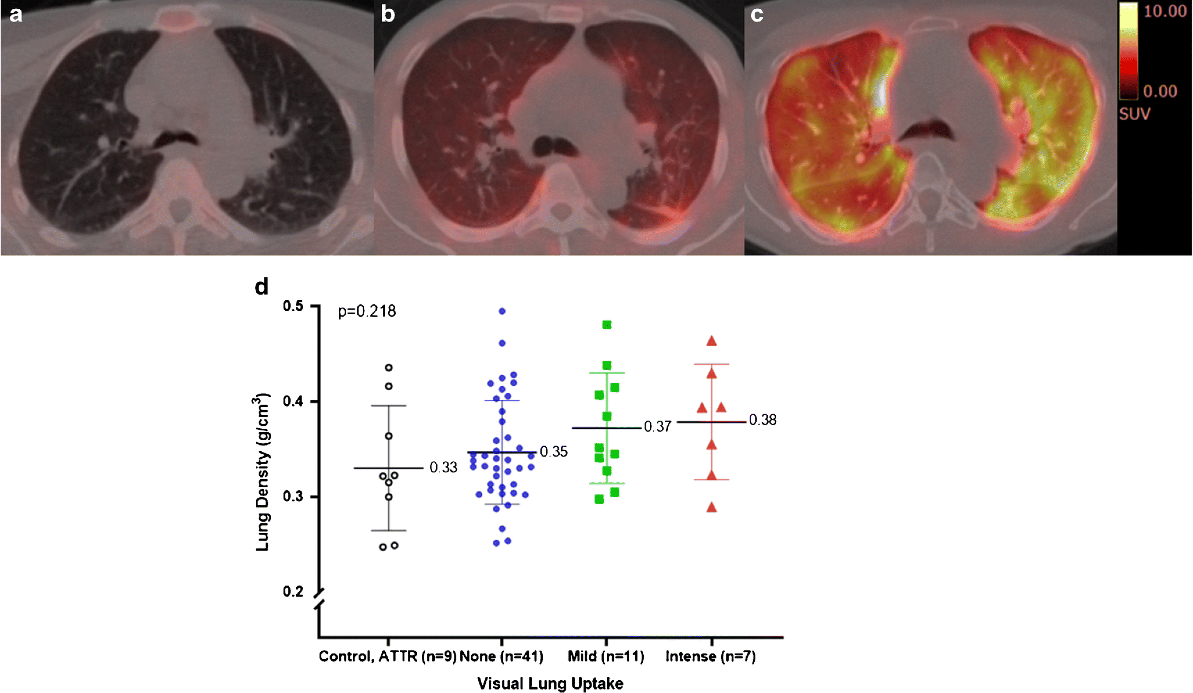
Axial fused [18F]florbetapir PET/CT delayed images (Top, a-c) from three subjects with no significant uptake (a), diffuse mild uptake (b) and diffuse intense uptake (c). Their left ventricular ejection fractions were 47%, 36% and 62% respectively. CT appearance of the lung parenchyma was unremarkable in these subjects.
CT average lung densities did not differ among subjects with no significant uptake, mild diffuse uptake, intense homogeneous uptake and the control group (bottom, d). The mean and standard deviation are shown. Each marker corresponds with a single subject