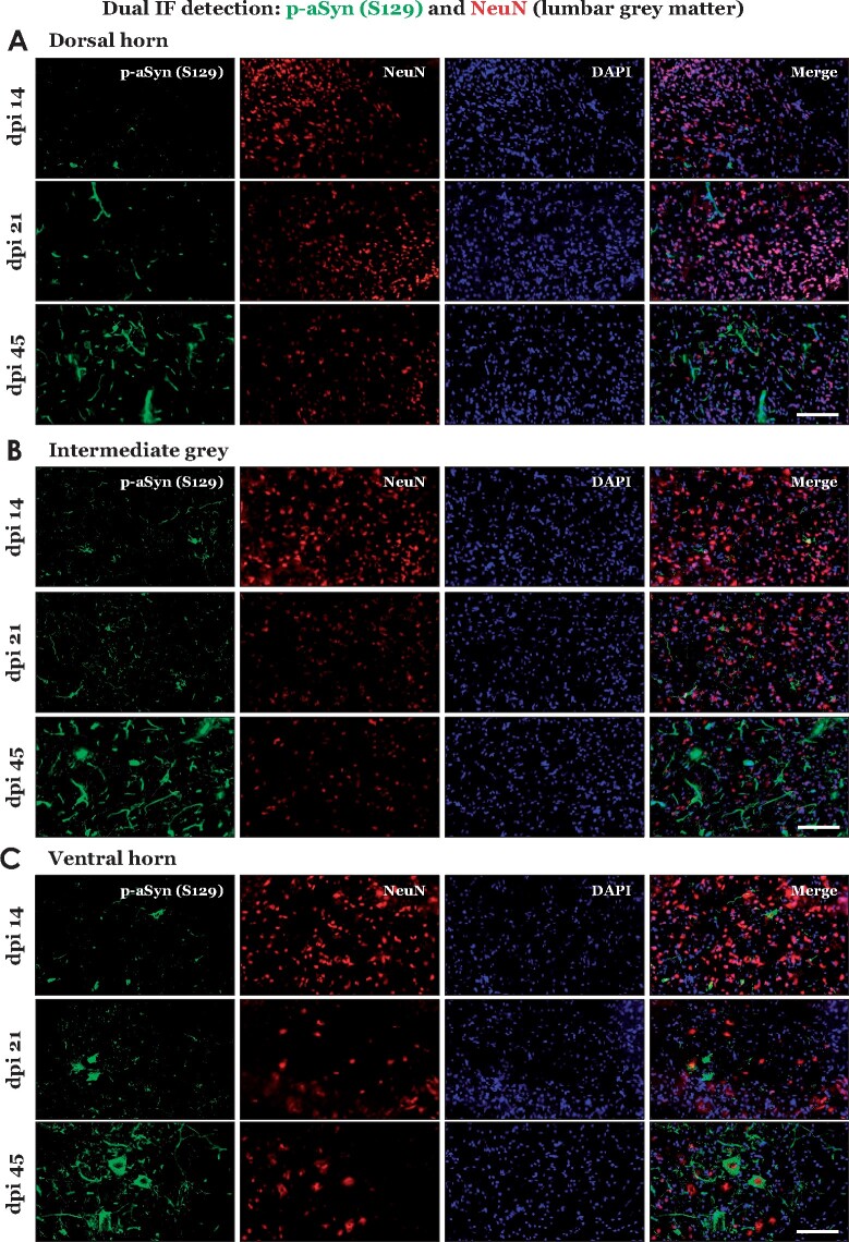
Figure 2.
Immunofluorescence detection of phospho-alpha synuclein (p-aSyn, S129) in the lumbar spinal grey matter of PFF aSyn injected M83+\+ mice. (A–C) Representative images showing dual immunofluorescence detection of p-aSyn (S129, in green) and neuronal nuclei marker (NeuN, in red) in lumbar grey matter of PFF aSyn injected cohort: dorsal horn (A), intermediate grey (B) and ventral horn (C). (dpi, days post-injection; 40× magnified views, scale bar = 50 µm). Also see Supplementary Fig. 1B, for representative images from the PBS injected cohort- dpi 45. Primary antibodies in A–C: p-aSyn (S129)- 11A5 and NeuN- A60.

