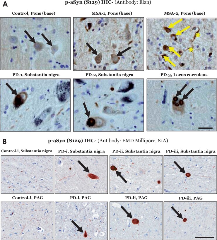
Figure 5.
Immunostaining of phospho-alpha synuclein (p-aSyn, S129) in post-mortem human brain sections. (A) Representative images showing p-aSyn (S129) immunostaining by the Elan antibody in a control (base of pons- basis pontis), two multiple system atrophy cases (MSA, #1–2, Supplementary Table S1; base of pons) and three Parkinson disease cases (#1–3, Supplementary Table S1; substantia nigra and locus coeruleus). The black arrows indicate neuronal p-aSyn (S129) inclusions in both Parkinson disease and MSA cases. In MSA-2, glial p-aSyn (S129) inclusions are indicated by the yellow arrows, while the fibre tract staining is indicated by the yellow asterisks (scale bar = 20 µm). (B) Representative images showing p-aSyn (S129) immunostaining by the EMD Millipore antibody 81A in the midbrain substantia nigra and periaqueductal grey-PAG of a control (#i, Supplementary Table S2) and three Parkinson disease cases (#i–iii, Supplementary Table S2). The black arrows indicate Lewy body pathology in Parkinson disease (scale bar = 100 µm). Also see Supplementary Fig. 6.

