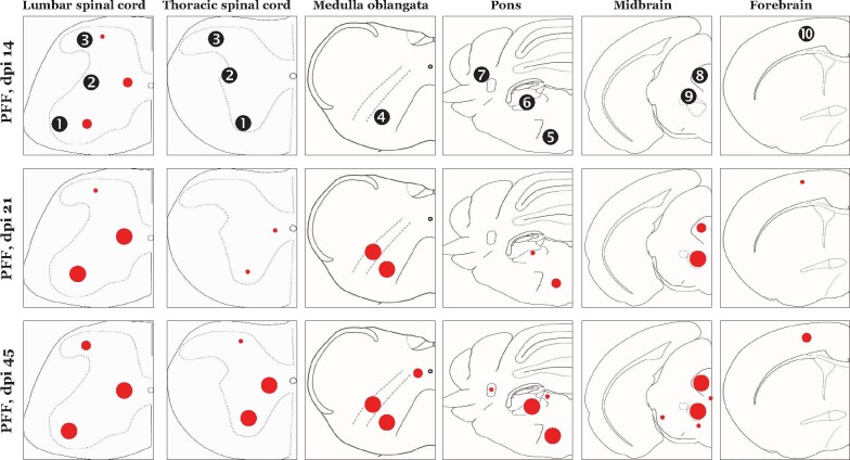
Figure 6.
Composite schematic depiction of the early stage phospho-alpha synuclein (p-aSyn, S129) immunodetection in the CNS of PFF injected M83+/+ mice. Average number of p-aSyn (S129) immunopositive cells in the regions is indicated by the red circles (small circle, 1–10 cells/mm2; medium circle, 10–30 cells/mm2; large circle, >30 positive cells/mm2). Regions shown: (Spinal cord) ventral horn, intermediate grey, dorsal horn; (Medulla oblangata) MdV, medullary reticular nuclei; (Pons) gigantocellualr nuclei, vestibular nuclei, cerebellar nuclei; (Midbrain) periaqueductal grey, ed nucleus; (Forebrain) motor cortex [dpi, days post-intramuscular injection with preformed fibrililar (PFF) aSyn; Structures are not drawn to scale]. Also see Figs. 1–3 and Supplementary Fig. 3.

