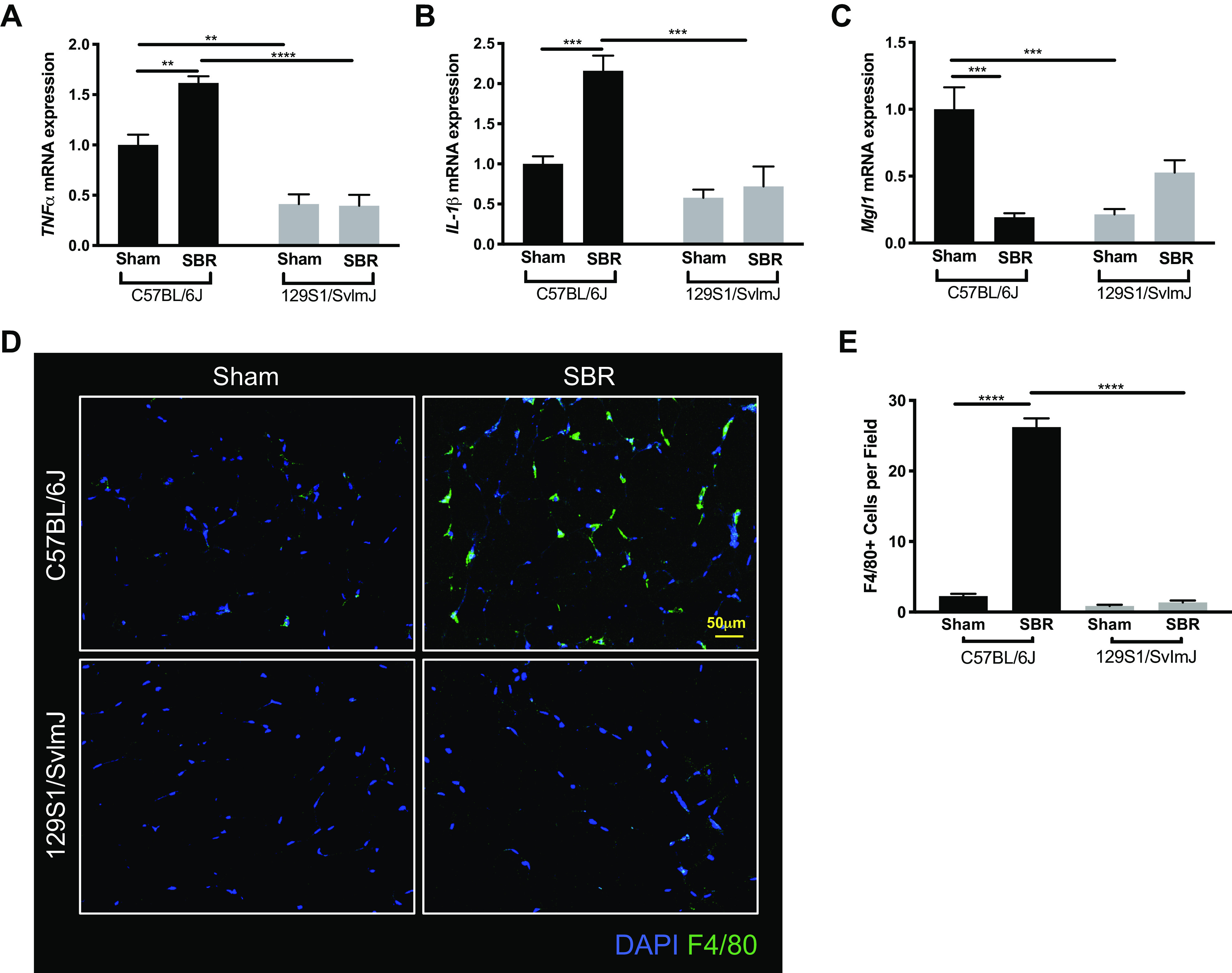
Figure 6.

mRNA expression levels of tumor necrosis factor α (TNFα) (A), interleukin 1-β (IL-1β) (B), and mitochondrial genome integrity 1 (MgI1) (C) in white adipose tissue from sham (n = 5, 4) and small bowel resection (SBR) (n = 5, 4) operated C57BL/6J and 129S1/SvImJ mice, respectively. D: representative images of F4/80-stained white adipose tissue. E: quantification of F4/80 staining for visceral white adipose tissue macrophages in sham (n = 3, 3) and SBR (n = 3, 3) mice in C57BL/6J and 129S1/SvImJ lines, respectively. ****P < 0.0001, ***P < 0.001, **P < 0.005.
